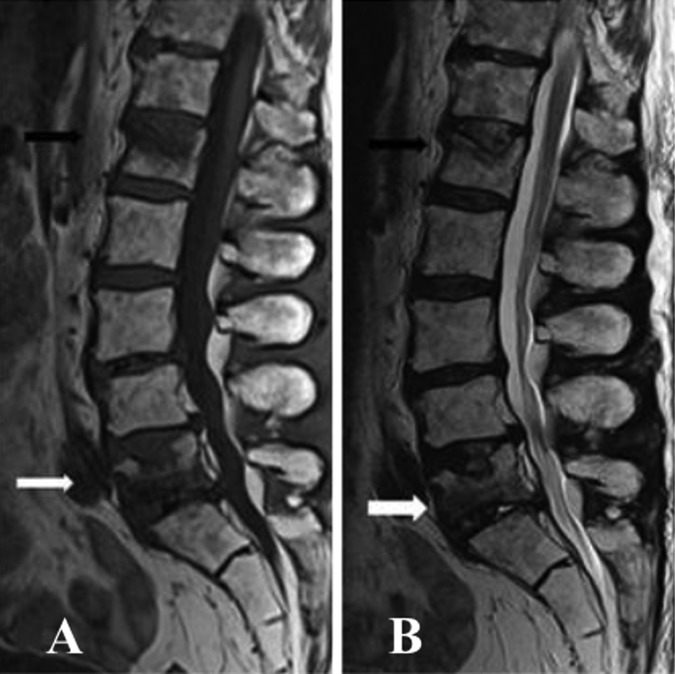
Figure 2.
(A and B) Sagittal T2 and T1 images of the lumbar spine demonstrated increased signals throughout the lumbar vertebrae in keeping with postradiotherapy change. Mild to moderate reduction in height of L1 vertebral body (white arrow) and L5 vertebral body (black arrow) with superior end-plate involvement would be the result of old compression fractures.

