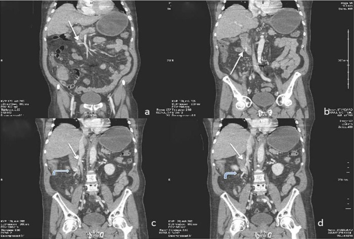
Figure 2.
Coronal reconstructed CT images through the second and third portions of the duodenum. (2A) White arrow better delineates varix originating from the superior mesenteric vein. (2B). Varix embedded in the lateral duodenal wall. Curved arrows in (2C and D) demonstrate the circuitous route the varix takes inferiorly ultimately draining into the posterior, lateral aspect of the inferior vena cava.

