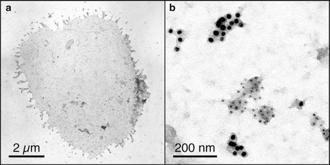
Fig. 1.
(a) Whole plasma membrane sheet from an activated T cell bound to an EM grid coated with stimulatory ligands (peptide-MHC II and co-stimulatory B7.1 molecules). (b) Magnified area of a plasma membrane sheet from a quiescent T cell bound to an EM grid coated with poly-l-lysine. Ectopically expressed myristoylated “non-raft” and myristoylated + palmitoylated “raft” markers are labeled with 5 and 10 nm gold particles, respectively.

