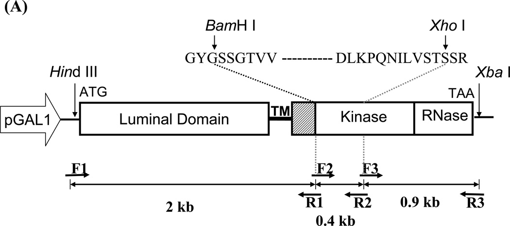
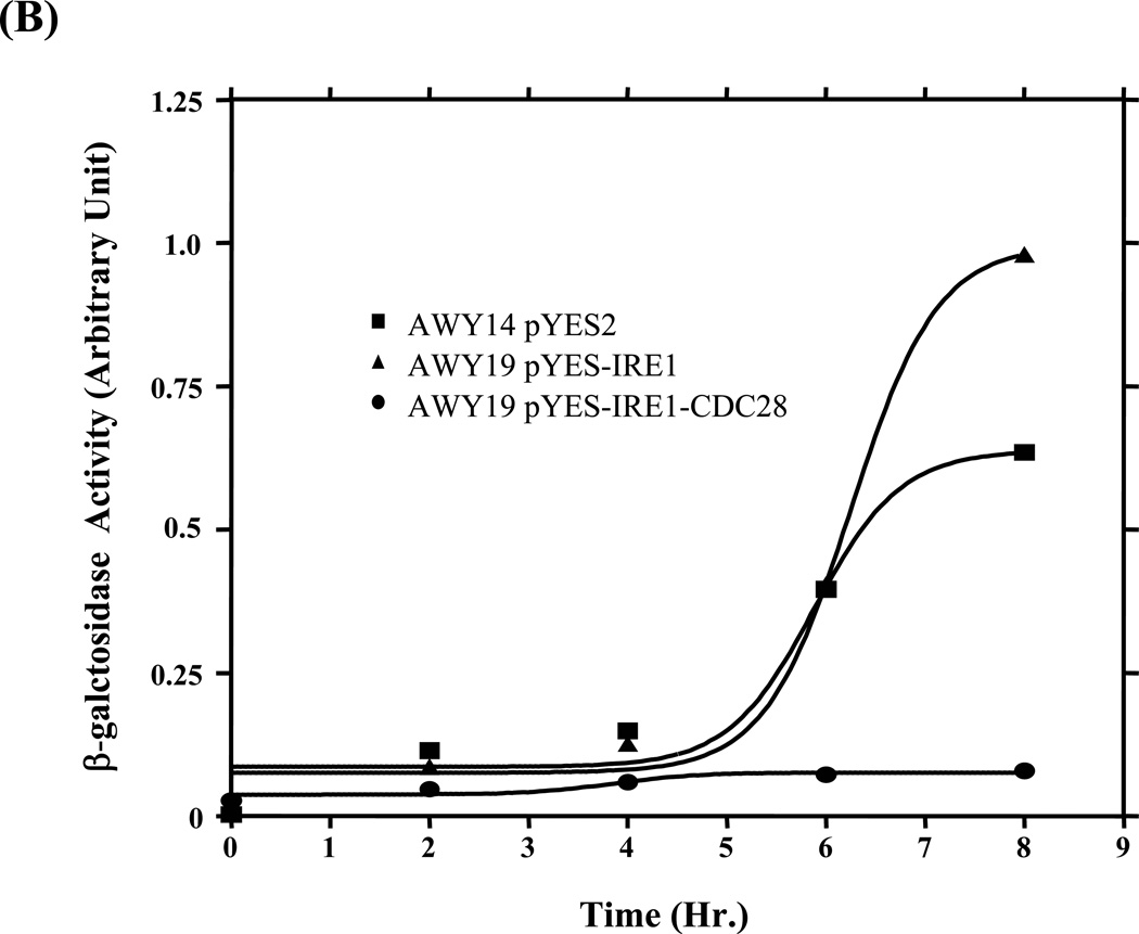
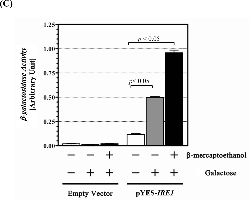
Figure 1. UPR restoration by inducible Ire1.
(A) Construction of the inducible Ire1 expression plasmid. The S. cerevisiae IRE1 gene was PCR amplified as 3 consecutive fragments then cloned under GAL 1 promoter in pYES-2 plasmid. BamH I and Xho I restriction sites (indicate by arrows) were introduced into the kinase subdomains I and VI, respectively. Positions of primers and the approximate size of the amplified fragments are indicated. GYGSSGTVV and DLKPQNILVSTSSR are conserved amino acid sequences in subdomain I and VI used to design degenerate primers. (B) Measure of UPR reporter gene activity. Wild type (AWY14) or ire1 null (AWY19) strains with indicated plasmids were cultured uracil dropout containing 2% raffinose, 1% galactose and 10 mM β-mercaptoethanol. β-galactosidase activity was measured at indicated time. (C) The UPR activity of the recombinant Ire1 in the presence of absence of ER stress was determined (Mean ± SEM) (n=3). ** represent significant difference of the reporter (P< 0.05, n = 3) by one-way ANOVA test.


