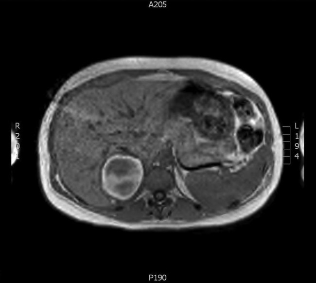
Figure 3.

T1-weighted imaging showing a decrease in volume of the adrenal mass, several months after diagnosis. The haematoma is becoming hyperintense from the peripheral rim, due to the oxidation and conversion of deoxyhaemoglobin to free methaemoglobin.
