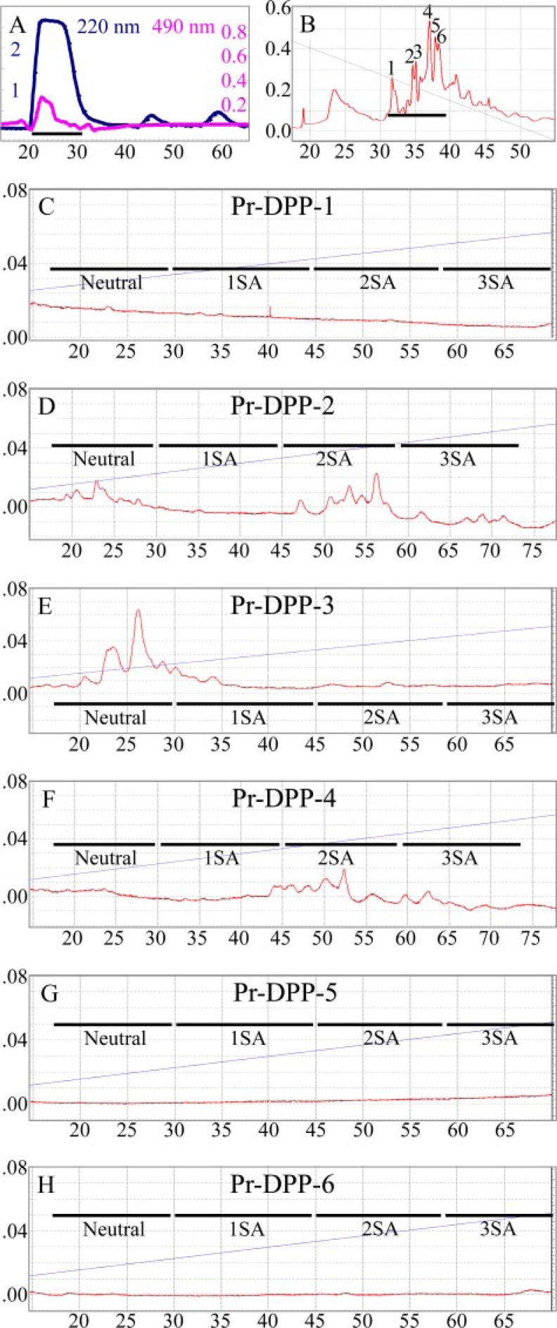
FIGURE 3.
Identification of two N-glycosylation sites in DPP. A, size exclusion chromatogram of pronase digestion products of DPP with absorbance monitored at 220 nm (blue line). Aliquots were analyzed for glycosylation by measuring the absorbance at 490 nm (magenta) after performing the phenol-sulfuric acid assay. The fraction applied to the next column is underlined. B, chromatogram of glycosylation-positive fraction (peak 1 in A) divided into 6 parts by hydrophilic interaction-HPLC. The 6 Pr-DPPs were digested with glycopeptidase A, and the released N-glycosylations were fluorescently labeled. C–H, chromatograms of labeled N-glycans on NP-HPLC and monitored with an excitation wavelength of 230 nm and an emission wavelength of 420 nm. No N-glycans were released from the Pr-DPP fraction 1 (C), 5 (G), or 6 (H). N-glycans were eluted in order of the number of sialic acids (SA) indicated as Neutral, 1SA, 2SA, and 3SA (34). The N-glycan for Pr-DPP-3 (E) has no SA, while those for Pr-DPP-2 (D) and Pr-DPP-4 (F) have two sialic acids.

