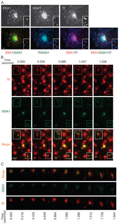
Figure 3. G-GGA/clathrin structures contain endocytosed Tf.

A) COS-1 cells expressing YFP-GGA1 were incubated with Alexa568 Tf for 1 hour at 37°C, then fixed and immunostained with anti-EEA1 antibody. Almost all the peripheral GGA1 signals overlap with Tf signal (arrows), but the majority of peripheral GGA1 structures do not contain EEA1. Inset shows an expanded view of boxed region. Bar, 10 μm. B) COS-1 cells expressing YFP-GGA1 were incubated with Alexa568 Tf for 15 min at 37°C and images of both GGA1 and Tf were simultaneously acquired using 36-millisecond exposures (Video S6). Rapid movements of G-GGA/clathrin structures along with Tf (boxed regions) are presented in a series of frames from Video S6. Bar, 1 μm. C) G-GGA buds decorate a rapidly moving Tf structure. Panel shows a montage of a series of frames from the boxed region (4 × 3.5 μm) of Video S7.
