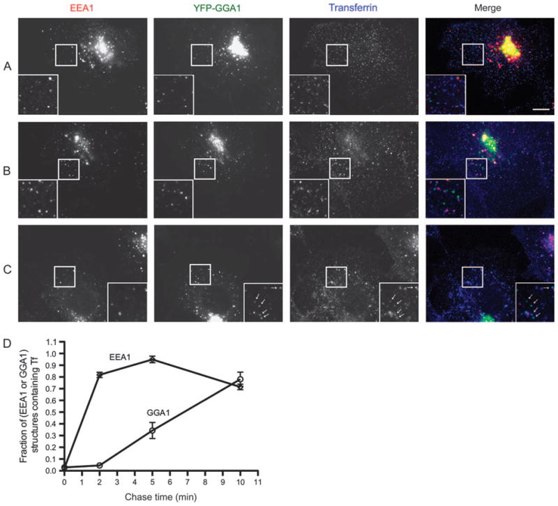
Figure 4. G-GGA/clathrin are post-sorting endosomal structures.

Cells transfected with YFP-GGA1 were pulse-labeled with Alexa647 Tf at 23°C for 1.5 min, chased for 0, 2 or 10 min, fixed and stained with EEA1 antibody. Inset shows an expanded view of boxed region. A) At start of chase, Tf labels only a few small peripheral EEA1-positive endosomes, and Tf is not detectable in GGA1 structures. Bar, 10 μm in all panels. B) After 2 min chase, almost all the EEA1 endosomes are labeled by Tf, while GGA1 structures still lack Tf. C) After 10 min chase, almost all the GGA1 structures contain Tf (arrows). D) Quantitative analysis of appearance of Tf in GGA1- and peripheral EEA1-positive structures (mean ± SEM). Ten regions from 10 cells at each time-point were analyzed.
