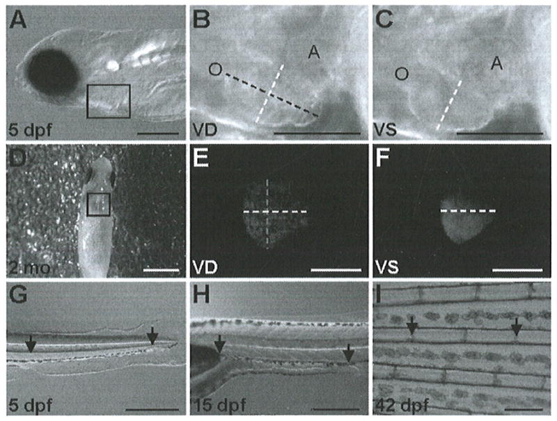
Fig. 1.

Orientations and measurement locations for shortening fraction, heart rate, and red blood cell flow rate assays. (a) For cardiac imaging, zebrafish up to 4 weeks old are placed on their right side in 3% methyl cellulose or E3, as shown with the 5-dpf zebrafish. (d) Fish older than 4 weeks are placed on a moist sponge, ventral-side up. (b, c, e and f) Maximum ventricular diastole (VD) and ventricular systole (VS) are shown, with the width depicted as a white dashed line and the length as a black or gray dashed line. Outflow tract (O) and atrium (A) are labeled in images (b, c). For the red blood cell flow rate assay, (g) 5-dpf, (h) 15- and 21 -dpf, and (i) 6-week fish are placed horizontally on their right side. Arrows refer to specific locations used for starting and stopping the stopwatch. Scale bars for (a–i) are 0.25, 0.125, 0.125, 3, 1, 1, 0.5, 0.5, and 0.5 mm, respectively. (Images (g–i) are reproduced from (21)).
