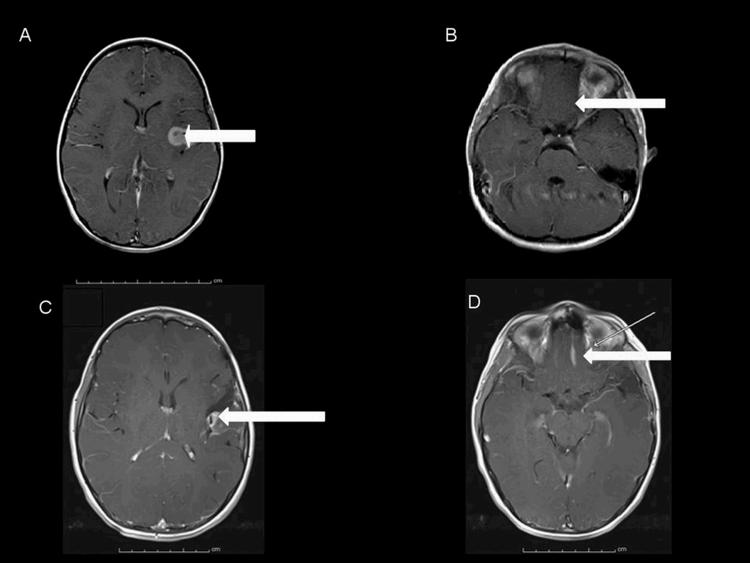
Figure 1.
Axial MRI studies obtained with contrast, in 2005, showing an enhancing lesion in the left sylvian fissure (A) and no obvious lesion in the left gyrus rectus (B). Axial postcontrast MRI studies obtained in 2009 showing a partially resected left sylvian fissure lesion (C) and a newly apparent and distinct lesion in the left gyrus rectus (D). The arrows designate the lesions (or in the case of panel B, the lack thereof).

