
Figure 2. PHB as an Intracellular Effector of TGF-β Signaling.
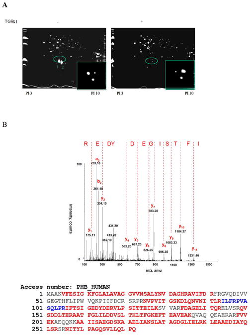


A. TGF-β leads to PHB dephosphorylation in prostate cancer cells. Cells were treated with TGF-β (12hrs) and soluble protein samples were resolved by 2D-gel electrophoresis and visualized with Coomassie blue. B. Differential protein spots circled, were sliced and sequenced by HPLC-MS, Spot 1 sequence coverage 71%; Spot 2 sequence coverage 75% Peptides highlighted in red were identified in both spots 1 and 2; peptides highlighted in blue were identified in spot 2. C. Protein samples (as above) were resolved through 2D-gel electrophoresis and transferred to PVDF membrane. Membranes were exposed to PHB and ERK1/2 antibody; the arrows indicate the protein PI shift. D. TGF-β activates Smad-dependent signaling. PC-3 cells were cultured in CCS medium, and after treatment with TGF-β, subcellular fractions or whole cell lysates were subjected to Western blotting for analysis of expression of Smads (Smad2/3 and Smad 4) signaling effectors. E. Raf is an upstream effector of PHB. PC-3 PHB shRNA transfectants (and non-silencing vector controls), were exposed to TGF-β (5ng/ml) for 1hr, and 12hrs. A marked increase in phosphorylation of both Raf and Erk (but not Akt) was detected within 1hr of TGF-β treatment. F. PC-3 shPHB transfectants (cultured in CCS medium), were treated with TGF-β and/or the Erk kinase inhibitor PD98059 (25μM) or UO126 (10μM). Phosphorylation of Raf protein was determined by fluoresence microscopy and Western blotting (upper and lower panel respectively).