
Figure 4. PHB Regulates Inner Mitochondrial Membrane Permeability in TGF-β Apoptotic Signaling downstream of Smad (Apoptosis) and MAPK.

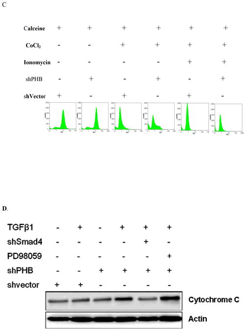

A. Calcein uptake by mitochondria in human prostate cancer cells; (staining with Calcein AM and MitoTracker Red). B. Decreased calcein signal correlates with increased CoCl2 concentration. Knockdown shPHB or sh vector transfected PC-3 cells were incubated with Calcein AM (100nM) and CoCl2 (15mins at 37°C) and events were detected as described as above. Data shown are the mean values from three independent experiments. C. Calcein AM loading assay. Stable transfectants shPHB or shNeg (106 cells) were incubated with 100nM Calcein AM, with or without CoCl2 (0.4mM) or ionomycin (0.5mM) (at 37°C; 15mim). Approximately 10,000 events were collected for flow cytometric analysis (PARTEC; 530nm Channel). D. Stable transfectants of PC-3 cells expressing the shVector, shPHB, shPHB, and transiently transfected with Smad4 siRNA or treated with PD98059, were exposed to TGF-β (5ng/ml); Soluble cytosolic fractions were subjected to Western blotting to examine mitochondrial cytochrome C release. E. Smad4 siRNA transfected PC-3 cells or mock-transfected control cells were treated with TGF-β for the indicated time periods with or without PD98059 or U0136, followed by Calcein AM loading assay; the values shown are the mean from three independent experiments performed in duplicate (p<0.05).