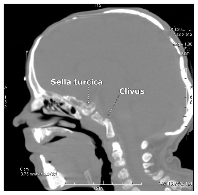
Figure 4.
Sagittal reformatted CT scan in a 15-year-old adult patient with cleidocranial dysplasia showed defective ossification of the sella turcica and the clivus. In addition, it showed that the entirety of the skull base and most of the sphenoid bone and the clivus are cartilaginous, while the remainder of the cranial vault underwent defective membranous ossification. The odontoid is hypoplastic, and there was agenesis of the anterior arch of the atlas. Also note the severe ossification defects of the anterior and the posterior fontanels respectively.

