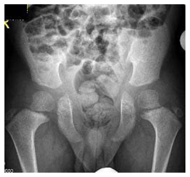
Figure 9.
AP radiograph of the pelvis in a 9-year-old patient with cleidocranial dysplasia. Note that the pelvis showed a combination of delayed ossification of the ischium and pubis. In addition, there was maldevelopment of the capital femoral epiphyses, and both were separated from the metaphyses with upward and medial inclination. Note the gap between the epiphyses and the irregular metaphyses respectively.

