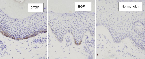
Figure 2.

On the 28th day, the fibroblast cytoplasm of the control group was not stained, while that in the two therapeutic groups showed a positive result. The positive cells were distributed not only in the papillary and subdermis, but also in some parts of epidermal layer of the healing wound. The epithelial layers in the rhEGF group were more than that in the rhβFGF group; the base membrane and the papillae of corium were evident at 28 days after treatment of rhEGF, that of rhβFGF group were blurring. (in situ hybridization × 400).
