Figure 6.
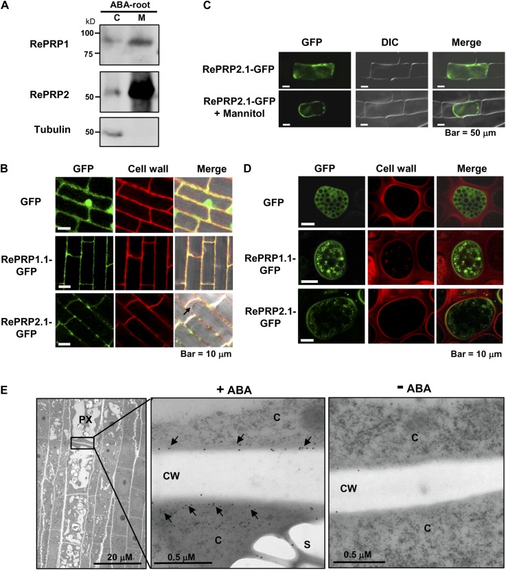
RePRP proteins are localized to the plasma membrane. A, Cytosolic (C) and membrane (M) proteins were extracted from 20 μm ABA-treated rice roots. RePRP1 and RePRP2 were detected mainly in the membrane fraction by western-blot analysis. Tubulin was used as a control for the cytosolic fraction. B, GFP and RePRP-GFP were stably expressed in rice. GFP was detected in cytosol and nucleus, whereas RePRP1.1-GFP and RePRP2.1-GFP were detected in cell boundary but not in cytosol (arrow) in root cells. Cell walls were stained by propidium iodide. C, GFP and RePRP2.1-GFP were transiently expressed in onion epidermal cells by particle bombardment-mediated transfection. RePRP2.1-GFP remained inside cells after plasmolysis by mannitol treatment. D, GFP and RePRP-GFP were transiently expressed in barley aleurone layers via particle bombardment. Cell wall was stained by propidium iodide. E, Localization of RePRPs in ABA-treated roots was detected by immunogold labeling with anti-RePRP2 antibody and observed by TEM. TEM images show a longitudinal section of the elongation region of ABA-treated young roots (left). Magnification of the boundary region between protoxylem (PX) cells shows gold particles (arrows) in cytoplasm (C) and the plasma membrane but not in cell walls (CW; middle). Only a few gold particles were detected in roots without ABA treatment (right).

