Figure 5.
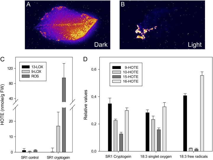
The main LPO processes in cryptogein-treated tobacco leaves in the light are caused by singlet oxygen. A and B, Autoluminescence imaging of detached leaves (treated as in Fig. 1, A and B). The necrotic areas show an emission of light attributed to lipid peroxidation processes. Photographs from one of two independent experiments are shown. C and D, Excised tobacco leaves treated with cryptogein and incubated for 48 h in continuous light (350 µmol m−2 s−1). Leaves developing HR symptoms were analyzed for lipid peroxidation processes. C, Quantification of lipid peroxidation mediated by 13-LOX, 9-LOX, or ROS and expressed as HOTE levels measured by HPLC-UV (Montillet et al., 2005). FW, Fresh weight. D, Specific HOTE distribution in ROS-mediated lipid peroxidation of elicited SR1 leaves, as measured by HPLC-tandem mass spectrometry (Triantaphylidès et al., 2008). As a reference, the distribution of the 18:3 oxidation by methylene blue in the light (singlet oxygen) and by tert-butyl hydroperoxide (free radicals) is also shown. Results are means ± sd of three analyses carried out on material from three leaves.

