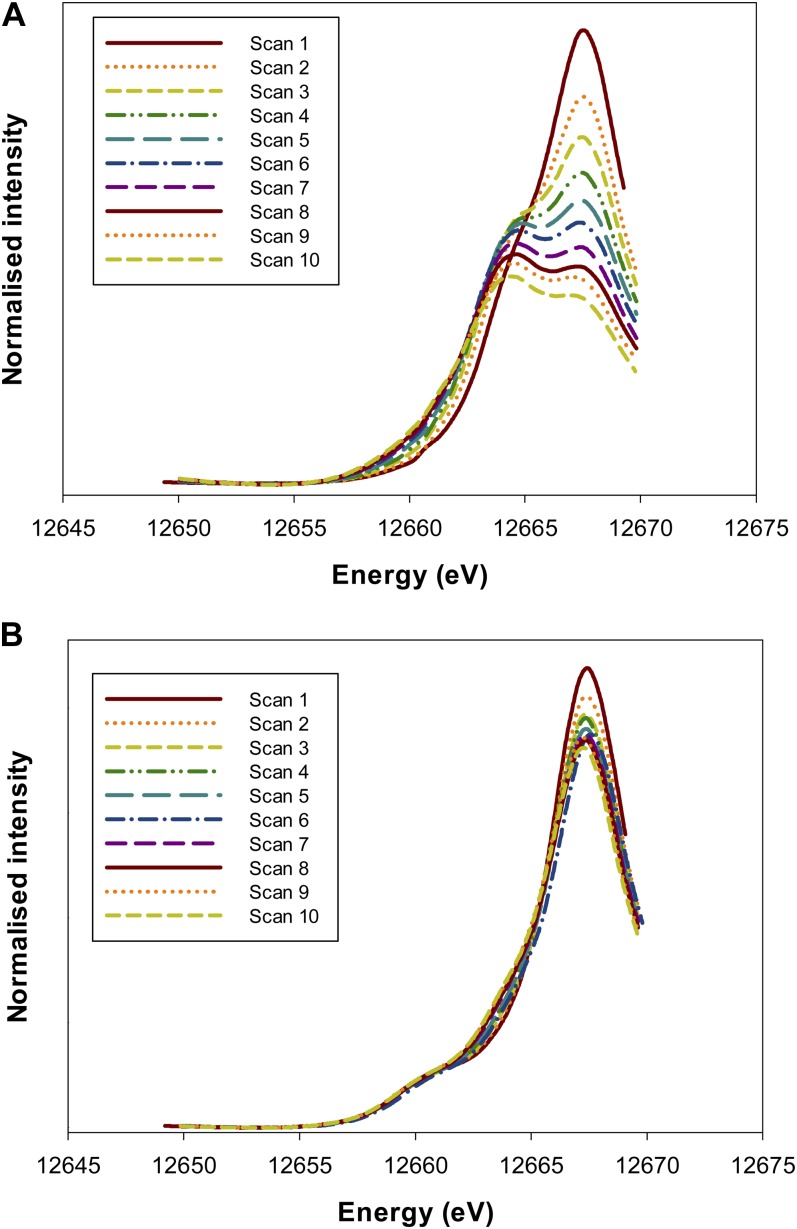
Figure 3.
Normalized Se Kα edge XANES spectra for an Na2SeO4 (Se[VI]) standard (A) and apices of cowpea roots exposed to 20 µm Se[VI] (B). The repeated rapid scans show the photoreduction of Se[VI] to Se[IV] induced by the x-ray beam. Also note that for the roots (B), the small peak at 12,661 eV does not appear to be an experimental artifact (i.e. it was observed on the first scan, and the magnitude of the peak did not increase progressively). [See online article for color version of this figure.]

