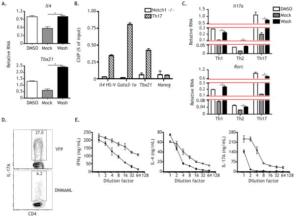
Figure 4. Notch simultaneously orchestrates multiple Th cell programs.
WT CD4+ T cells were stimulated as described under Th17 culture conditions. (A) After 24 hours, cells were treated with either DMSO or GSI for 20 hours and then subjected to a GSI washout assay. Cells cultured in DMSO were replated in DMSO and cells cultured in GSI were either replated in GSI (Mock) or in DMSO (Washout) for 4 hrs. RNA was then harvested and analyzed by qPCR. (B) After 2 days, cells were fixed and ChIP was performed using anti-Notch1 antibody, with Nanog serving as an internal negative control. Anti-Notch1 ChIP was performed on CD4-Cre x Notch1FL/FL CD4+ T cells as an experimental and antibody negative control. (C) A GSI washout assay was performed as above on activated CD4+ T cells cultured under either Th1, Th2, or Th17 polarizing conditions. At the end of the assay (48 hrs), RNA was harvested and analyzed by qPCR. (D) Naive CD4+ T cells were FACS sorted from Tat-Cre treated YFPFL/FL or DNMAMLFL/FL CD4+ T cells. Cells were then stimulated as above, and cultured under Th17 conditions. After 3 days, cytokine production was measured by intracellular FACS. (E) WT CD4+ T cells were stimulated as described and then cultured in a titration series of two-fold serially diluted Th1 (left panel), Th2 (middle panel), or Th17 (right panel) conditioned media, in the presence of either DMSO or GSI. After 5 days, cells were restimulated and supernatants were analyzed by ELISA two days later. *, P < 0.05. Data are represented as mean +/− SEM. See also Table S1 and Table S2.

