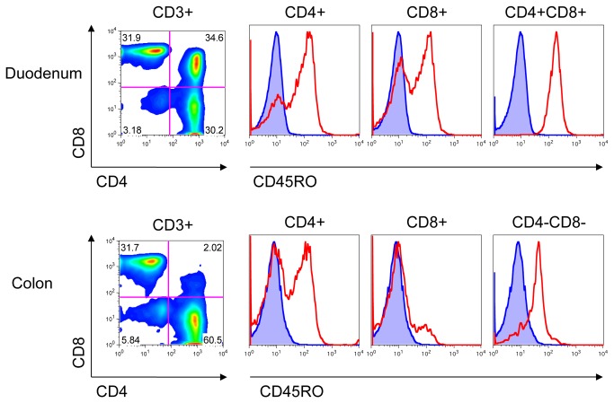
Figure 7. CD45RO expression on T-cells in the gut.
Lymphocytes were isolated from GALT of colon and duodenum of a SHIV-infected pigtailed macaque at time of necropsy and stained with Abs to CD3, CD4, CD8, and CD45RO. Cells were analyzed by FACS. Lymphocytes were gated by light scatter, and then on CD3+ cells. Cells were further gated on CD4 and CD8. Expression of CD45RO is shown in red. Unstained cells are shown in blue. Data representative of n=5 macaques.

