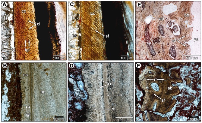
Figure 7. Comparisons of the periodontal tissues between diadectids and a fossil horse.
A: longitudinal section of cellular cementum in a molariform tooth of Equus sp. (ROM 33036) under normal light. B: longitudinal section of the acellular and cellular cementum of a diadectid molariform tooth (TMM 43628-3). Image was taken using an oblique illumination slider to highlight the incremental growth lines in the cellular cementum. C: longitudinal section of cellular cementum in Equus sp. (ROM 33036) under cross-polarized light. Note the extensive network of parallel Sharpey's fibers that mark the insertions of the periodontal ligament. D: closeup of the acellular and cellular cementum of a diadectid tooth (TMM 43628-3). Note the presence of a network of parallel Sharpey's fibers that mark the insertions of the periodontal ligament. E: closeup of the alveolar bone of Equus sp. (ROM 33036) in cross-section. Note the presence of Sharpey's fibers in the alveolar bone layers that border the periodontal space. F: closeup of the alveolar bone of a diadectid (TMM 43628-3) in cross-section. Note the presence of dense networks of Sharpey's fibers in successive layers of alveolar bone. A reversal line separates each layer of alveolar bone. ab, alveolar bone; ac, acellular cementum; bb, bundle bone layer of the alveolar bone; cc, cellular cementum; cl, cementocyte lacunae; igl, incremental growth lines in the cementum; ps, periodontal space; rl, reversal line; sf, Sharpey's fibers.

