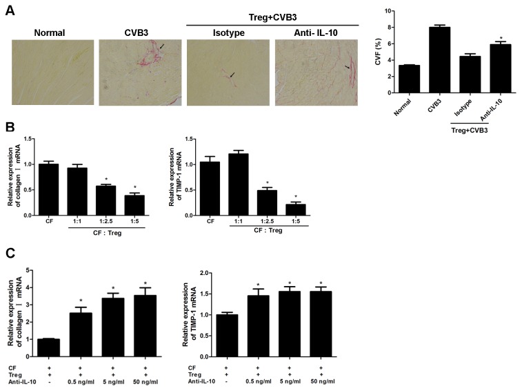
Figure 5. IL-10 played an important role in Tregs-mediated inhibition of cardiac fibrosis.
(A) Groups of 10 mice were adoptively transferred with 1×10⁶ Tregs one day before CVB3 infection. 1 hr after CVB3 infection, mice were injected with 100 μg of anti-IL-10 neutralizing mAb per mouse twice a week for 4 weeks. Representative Picrosirius-red stained heart sections 4 weeks after CVB3 infection (magnification: 200×) and the collagen volume fraction (CVF) was shown. Arrows indicate myocardial interstitial and perivascular fibrosis. Data represent the mean ± SEM. *, Individual experiments were performed three times. P<0.05 compared to mice given isotype Ab after Treg transfer. (B) RT-PCR measurement of cardiac fibroblast gene expression levels of collagen I and TIMP-1 24 hours after isolated Tregs were co-cultured with cardiac fibroblasts (CFs) at different cell ratios (1:1, 1:2.5, 1:5). Data are from one representative experiment of three performed ones and represent as the mean ±SEM. *, P<0.05 compared to cardiac fibroblasts culture alone. (C) The relative expression of collagen I and TIMP-1 after anti-IL-10 mAb was added in the co-culture system. Data represent the mean ± SEM, representative of three independent experiments. *, P<0.05, compared to co-culture system with no IL-10.

