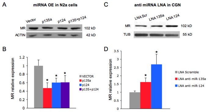
Figure 4. miR-135a and miR-124 regulate the expression of the endogenous MR protein.
(A) N2a cells were transfected with miR-135a (p135a) and miR-124(p124) overexpressing vectors as indicated. p135a+p124 corresponds to the transfection with an equimolar mixture of the two vectors. A representative blot is shown. (B) Western blot analysis of endogenous MR expression was quantified by densitometry, normalized to actin as loading control and expressed relatively to empty vector transfected cells. Data represent the mean from three biological samples and three technical replicates ± SE. *P < 0.05, **P < 0.05 (pairwise Student’s t-test). (C) Cerebellar granule neurons (6+4 DIV) were transfected with LNA antisense oligonucleotides or scramble LNA as negative control. A representative blot is shown. (D) Western blot analysis of endogenous MR expression was quantified by densitometry, normalized to tubulin and expressed relatively to scramble LNA transfected cells. Data represent the mean from four biological samples and three technical replicates ± SE. *P < 0.05, (pairwise Student’s t-test).

