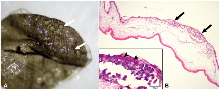
Figure 2. Biopsy of uropatagial membrane of Myotis myotis with acute inflammatory response against Geomyces destructans.
2A (sample HU 1): Dissection microscope image of punch biopsy with distinct circular lesions (arrows), where G. destructans had sloughed off during preparation. 2B: Histological cross section of 2A with multiple well-demarcated intraepidermal microabscesses (arrows). Hematoxylin-eosin staining. Inset: Hyphae of G. destructans invading the microabscesses (arrow heads). PAS staining.

