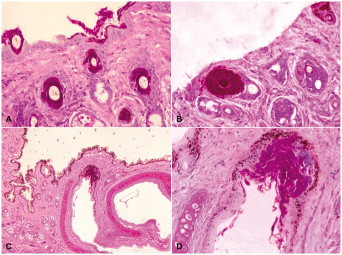
Figure 4. Skin of the muzzle of a Geomyces destructans infected Myotis daubentonii (sample NL 1).
4A: Multiple hair follicles with intraluminal colonization of G. destructans hyphae. 4B: Wall of a hair follicle replaced by marked growth of G. destructans hyphae. 4C+D: Cutaneous epithelium of one nare severely colonized by G. destructans with mild localized suppurative inflammation limited to the epithelial layer. PAS staining.

