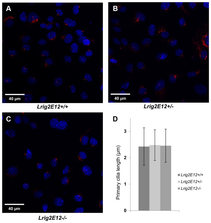
Figure 7. The length of the primary cilia in growth-arrested primary MEFs.
Wild-type, heterozygous, and Lrig2-deficient cells were serum starved for 24 hours followed by staining of primary cilia with antibodies against acetylated tubulin (red). The cell nuclei were counter-stained with DAPI (blue). Representative confocal immunofluorescence micrographs of: (A) wild-type (Lrig2E12+/+) cells, (B) heterozygous (Lrig2E12+/-) cells, and (C) Lrig2-defecient (Lrig2E12-/-) cells. (D) Quantification of primary cilia length. Shown are the means from three independent experiments including wild-type (n=8), heterozygous (n=9), and Lrig2-deficient (n=6) cell lines from three different litters, with standard deviation indicated by error bars. There were no differences observed in the abundance (data not shown) or the length of primary cilia in cells of different Lrig2 genotypes.

