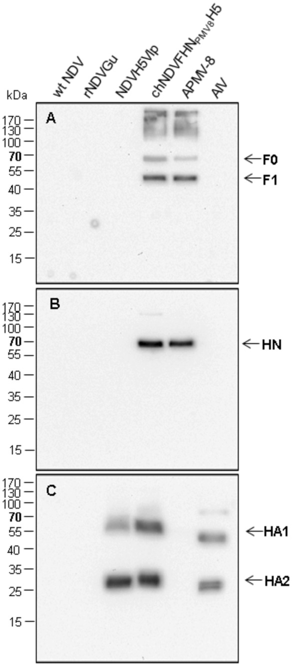
Figure 2. Protein expression and incorporation.

After Western blotting of purified virion lysates, envelope proteins were visualized by immunostaining with rabbit-α-APMV-8 F (A), rabbit-α-APMV-8 HN (B) and rabbit-α-AIV H5 (C). After incubation with the respective primary antibody, binding of peroxidase-conjugated species-specific secondary antibody was detected by chemiluminescence substrate (Pierce). Identified proteins are indicated on the right, molecular weights of marker proteins (PAGE Ruler™ Prestained Protein ladder (Fermentas)) are indicated on the left.
