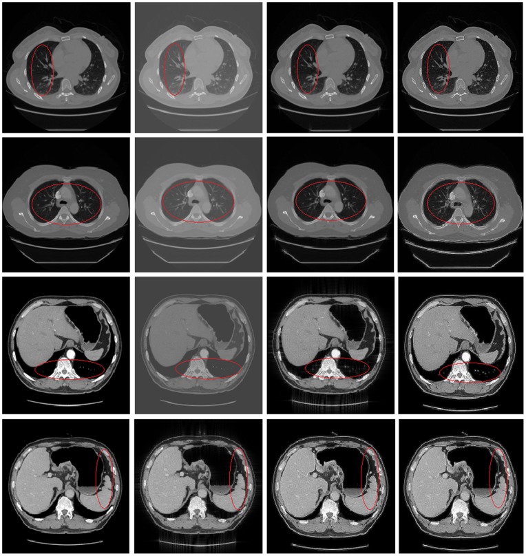
Figure 5. Shows comparisons of different methods.
From top to bottom: chest CT experiment, lung CT experiment, stomach CT (1) experiment, stomach CT (2) experiment. From left to right in each row: the original image, the recovery image using Wang's method, the recovery image using Hussien's method, the recovery image using our method. It needs to say, we select the RPCA model in chest CT and lung CT, and select the GoDec model in stomach CT. We can see that using our method to restore the medical CT image sequence can obtain satisfactory results, the recovery images can show high contrast, clear detail information and clear organ boundaries which are especially shown in the components circled with red ellipses in the images.

