
Figure 1.
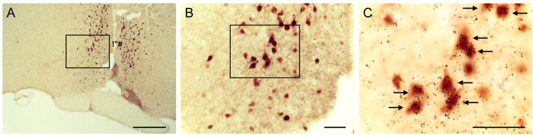
Co-expression of growth hormone secretagogue receptor (GHSR) mRNA and eGFP protein in the anteroventral periventricular/periventricular nucleus. Photomicrographs of a representative GHSR-eGFP mouse brain, sectioned coronally, at approximately 0.02 mm rostral to Bregma. Cells with eGFP-immunoreactivity have been stained with DAB and appear orange-brown in color (A–C). Those eGFP-immunoreactive cells that co-express GHSR mRNA have overlying black silver granules, and are indicated by arrows. Subsequent images on the right (B and C) are magnified images of the boxed areas in the images on the left (A). 3V, Third ventricle. Scale: 250 μm (A), 50 μm (B and C).