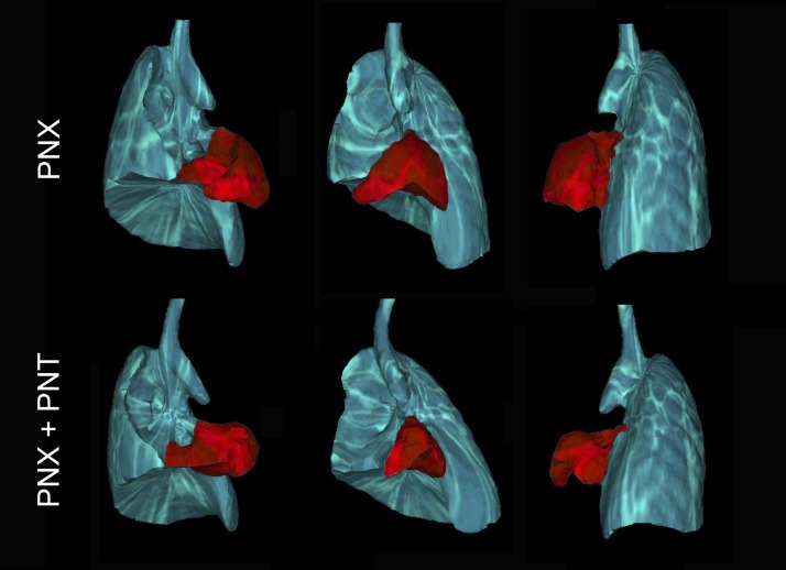
Fig. 4.
Respiratory-gated microCT scans demonstrating the anatomic configuration of the thorax at peak inspiration. Respiratory gating was used to obtain images at a comparable phase in the respiratory cycle. The lung is shown after PNX without (top) and with (bottom) PNT. The cardiac lobe is pseudocolored red for presentation. Cardiac lobe volumes, based on inspiratory-gated microCT scans, were 22% lower in mice after PNX + PNT (P < 0.05).

