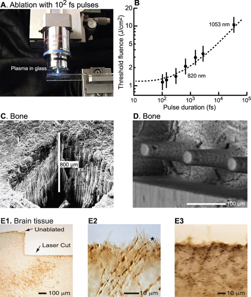
Figure 3. Plasma-mediated ablation of biological tissue with ultra-short laser pulses.

(A) Emitted light from the plasma bubble in glass melted by 100-fs pulses whose fluence is well above threshold. Adapted from [105]. (B) Plot of minimum fluence for ablation as a function of pulse width. The short 100 fs pulses have the minimum fluence. Adapted from [30]. (C) Scanning electron micrograph of a procine long bone cut in air. Adapted from [37]. (D) Scanning electron micrograph of patterned bone cut in air. Adapted from [38]. (E1) Bright-field image of immunostained surface of fresh brain tissue from rat, cut under saline. After completion of the optical ablation, the tissue was fixed, frozen, physically sectioned at a thickness of 25 μm, immunostained with anti-tyrosine hydroxylase, and visualized with diaminobenzadine precipitation. The brown regions correspond to immunostained axons and cell bodies. (E2) Tissue similar to that in panel G but imaged at high magnification to illustrate the cutting of individual axons (*). (E3) Immunoreactivity near an optically cut surface in unfixed neuronal tissue.
