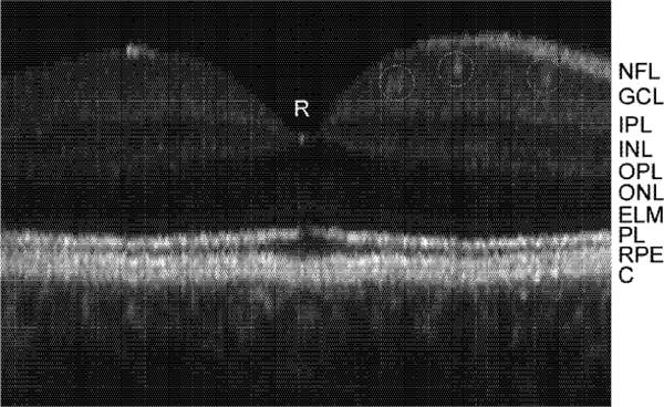
Fig. 8.

Magnified TOCT composite (24 frame) image of fovea with labeled retinal layers. Small retinal vessels are also identified with circles. NFL: nerve fiber layers, GCL: ganglion cell layer, IPL: inner plexiform layer, INL: inner nuclear layer, OPL: outer plexiform layer, ONL: outer nuclear layer, ELM: external limiting membrane, PL: photoreceptor layer (interface between inner and outer segments delineated by bright reflection), RPE: retinal pigment epithelium, C: choriocapillaris and choroid.
