Fig. 9.

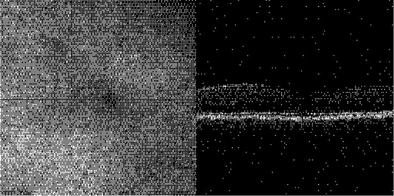
(3.5 Mb) En face (left) and cross-sectional scans (right) through the macular region Fovea is dark region in the center on the right and the region without overlying nerve fiber layer on the right.

(3.5 Mb) En face (left) and cross-sectional scans (right) through the macular region Fovea is dark region in the center on the right and the region without overlying nerve fiber layer on the right.