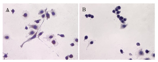
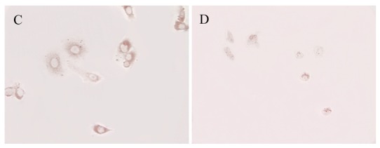
Figure 2.


Immunolocalization of ATIP in PC3 cells (A and C) with haematoxylin counter-stain (A) and without counter-stain (C); (B and D) serial cover slips pre-absorbed with ATIP antigen with (B) and without (D) haematoxylin counter-stain respectively. Images were taken at 400× magnification.
