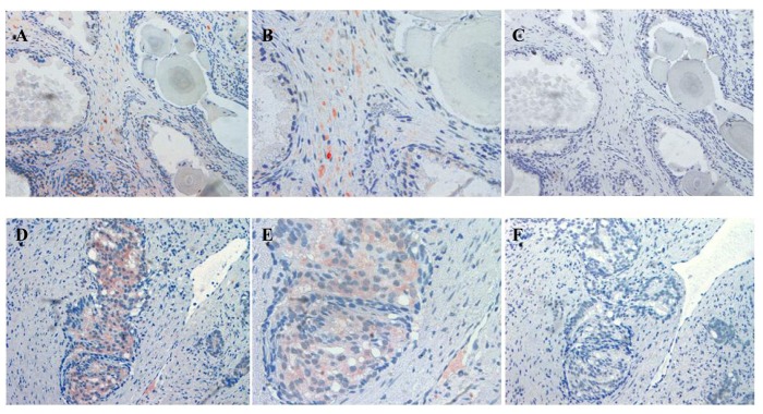
Figure 3.
Immunolocalization of ATIP staining in stroma surrounding BPH (A and B) and HGPIN (D and E) gland in paraffin embedded tissue sections. Serial sections showing an absence of staining when the ATIP antibody was pre-absorbed (C and F) with ATIP antigen. Images were taken at (A, C, D and F) 200× and (B and E) 400× magnification.

