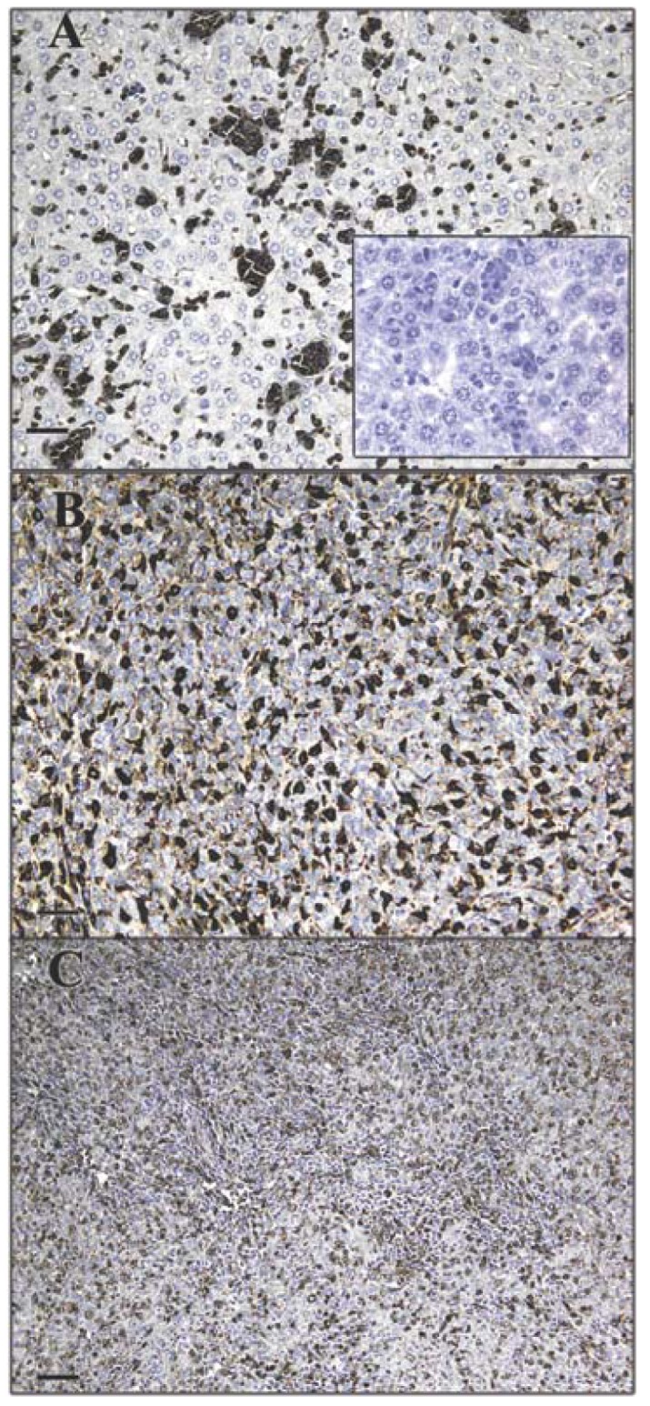
Figure 1.

Expression pattern of GS-I binding to tumor cells. Murine 4T-1 tumor transplants were labeled with 2.5 μg/mL GS-I. Metastatic tumor cells in the liver (A) and lung (B) demonstrate increased GS-I binding compared to cells within the primary tumor (C). 200× magnification, bars equal 40 μm. No staining is observed in sections stained with biotin alone (inset, A).
