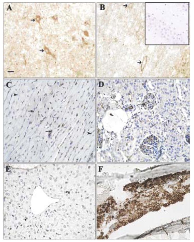
Figure 2.
Expression pattern of GS-I binding to normal cells. Tissues from a naïve control mouse were labeled with GS-I. (A) Brain: Neurons (arrows) were labeled in a cytoplasmic and membranous fashion; (B) Spinal cord: Neurons were labeled in a membranous pattern; (C) Heart: Endothelial cells in the interstitium and in small arteries (arrows) were labeled. Myofibers (arrowheads) were not stained; (D,E) Kidney, Liver: Endothelial cells (arrows) bound GS-I weakly to moderately. Hepatocytes and renal tubular and glomerular epithelial cells did not bind GS-I; (F) Bone marrow: Hematopoietic cells bound GS-I strongly in a membranous and cytoplasmic pattern; (A,B,C) 200× magnification, bar equals 40 μm. (D,E,F) 400× magnification, bar equals 20 μm. No staining was observed in sections stained with biotin alone (inset, B).

