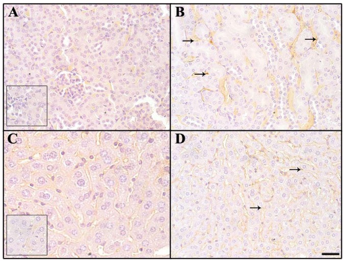
Figure 6.
Comparison of IgG and IgM binding of tissues of non-immunized mice. Normal tissues were immunostained with antibody against naturally deposited murine IgG (A,C) and IgM (B,D). IgM staining is more intense, with a membranous and punctate pattern on endothelial cells (arrows). No staining is present in negative controls labeled with goat immunoglobulin (inset A and inset C). Kidney (A,B) and liver (C,D), 400× magnification, bar equals 40 μm.

