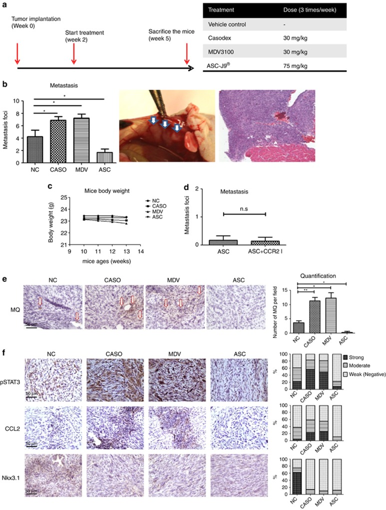
Figure 4.
Induction versus suppression of PCa metastases by Casodex, MDV3100, and ASC-J9. (a) Outline of the in vivo experiment. The TRAMP-C1 cells were used as in vivo mouse model to detect the tumor metastases in the transplanted nude mice. (b) Quantification of metastatic lesions in mice treated with different anti-A/AR compounds. After euthanizing the mice, the metastatic tumors were evaluated, and the numbers of metastatic foci in each mouse were quantified (left). The metastatic tumors observed on the diaphragm (middle) were confirmed by histology with HE staining (right). (c) Body weights of mice under anti-A/AR compounds treatment. The mice body weights were checked weekly starting from the first injection of compounds. (d) Quantification of metastatic lesions in mice treated with ASC-J9 alone or combined with CCR2 antagonist. (e) Migration of macrophages (arrows) into the primary tumor site. The paraffin-embedded tumor tissue sections were stained using antibody of macrophage marker F4/80 (left). The total numbers of macrophages (Mφ) in three randomly selected fields were quantified on the right. (f) Comparisons of STAT3/CCL2 signal in in vivo PCa tissue. The pSTAT3, CCL2, and Nkx3.1 expressions were detected by immunohistochemistry staining of the paraffin-embedded tissue sections ( × 400), the quantification results were shown on right. Error bars=mean±S.E.M. *P<0.05, **P<0.01. N.S., not significant

