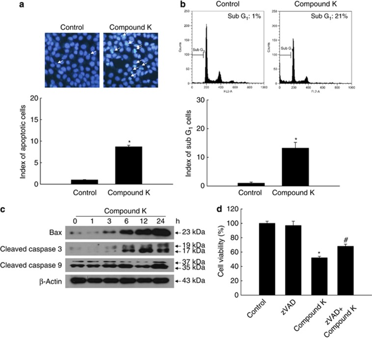
Figure 5.
Compound K induces apoptosis in HCT-116 cells. HCT-116 cells were treated with compound K (20 μg/ml) for 24 h. (a) Apoptotic body formation was observed after Hoechst 33342 staining and quantified. Apoptotic bodies are indicated by arrows. (b) The apoptotic sub-G1 DNA content was detected by flow cytometry after PI staining and quantified. (c) Bax, active (cleaved) caspase-3, and caspase-9 levels were assessed by western blot analysis. (d) Cells were treated with compound K (20 μg/ml) and/or Z-VAD-FMK (10 μM) for 24 h, and cell viability was assessed using the MTT assay. *Significantly different from control (P<0.05) and #significantly different from compound K-treated cells (P<0.05)

