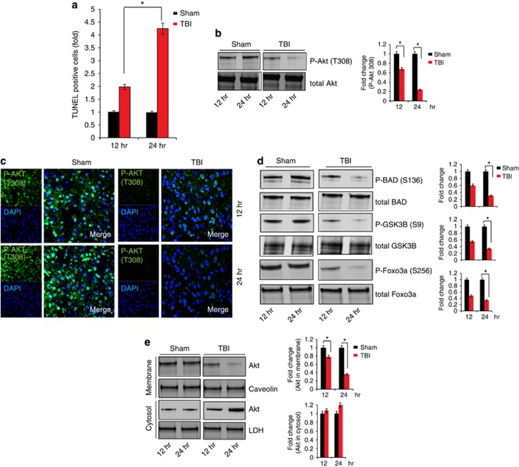
Figure 1.
Inactivation of Akt is associated with cell death following TBI. (a) TUNEL staining was done to identify cell death at 12 and 24 h after TBI. Quantitative analysis shows that TUNEL staining was increased more than twofold after 24 h post TBI in the pericontusional cortex. (b and c) Phosphorylation of Akt (T308) was determined by western blot and immunofluorescent microscopy. Changes in phosphorylation status of Akt (P-Akt T308) was measured quantitatively. (d) Phosphorylation of downstream proteins of Akt, such as GSK3B, Foxo3a and BAD was determined by western blot analysis 12 and 24 h post TBI. (e) Membrane and cytosolic fraction of Akt was determined in the cortex at 12 and 24 h post Sham or TBI in mice. Level of both cytosolic and membrane Akt was determined at 12 and 24 h post TBI quantitatively. *P<0.01, n=3, one-way ANOVA, mean±S.E.M.

