Figure 1.
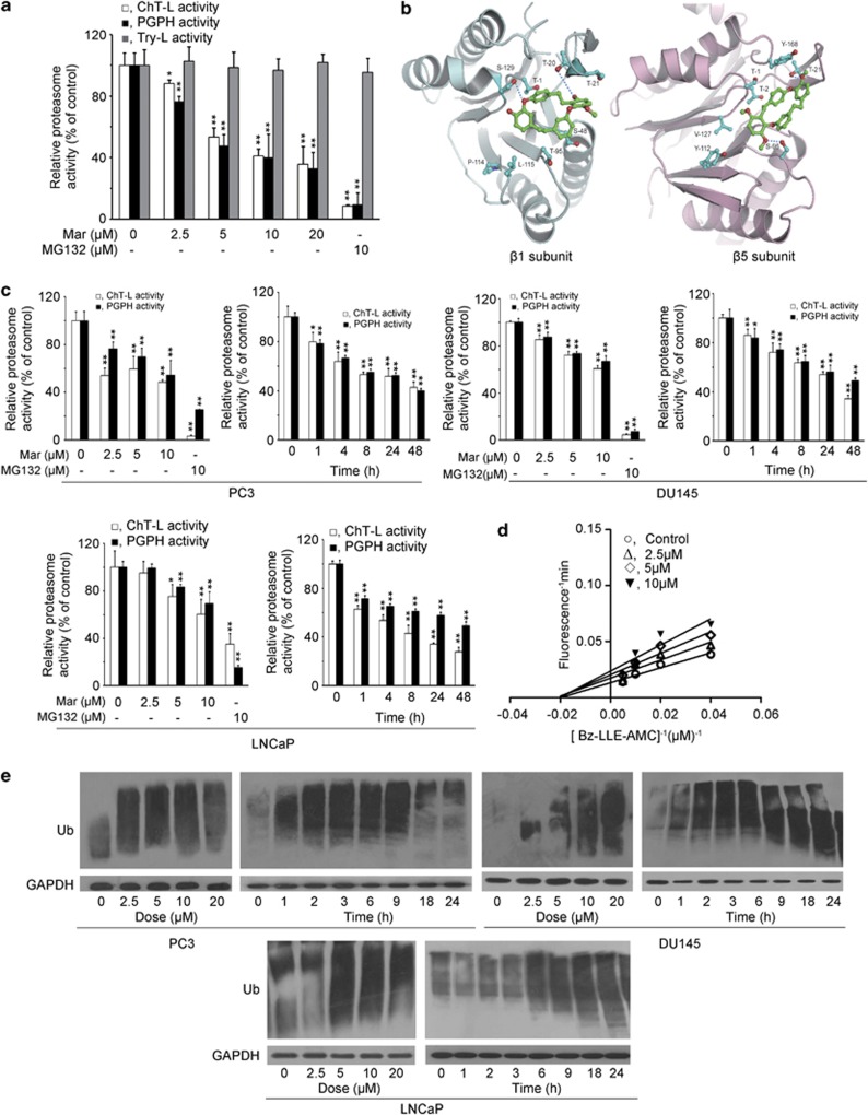
Mar inhibits ChT-L and PGPH activities of proteasome. (a) Purified human 20S proteasome was incubated with Mar. ChT-L, Try-L and PGPH activities were monitored with specific fluorescent substrates. Relative proteasome activity represented the percentage of fluorescence compared with the control. *P<0.05, **P<0.01, compared with the control (neither Mar nor MG132 addition). Data shown are means±S.D., n=3. (b) The stereo view of interactions between Mar molecule and the β1 (left panel) and β5 subunits (right panel) of proteasome. Green, Mar; cyan-blue, side residues of amino acids in β1 and β5 subunits. The residues interacting between Mar and β1 and β5 of 20S proteasome are labeled in red. The sequence alignment of β1 between the model and human proteasome is shown below, and the interacting residues are shown in bold: model 1TTLAFR7 16DSRTTTGAYIANRVTDKLTQHDTIWCCRSGSAA53; human 35TTLAFK41 51DSRTTTGSYIANRVTDKLTPIHDRIFCCRSGSAA84; model 94LTAGLIIAGWDERHGGQVYSIPL115 124YAIGGSGS131; human 128LMAGIIIAGWDPQEGGQVYSVPM150 159 FAIGGSGS166. The sequence alignment of β5 between the model and human proteasome is shown below, and the interacting residues are shown in bold: model 1TTTLAFRFQGGIIVAVDSRATAGNW25 44TMAGGAA50 93AGLSMGT99; human 1TTTLAFKFRHGVIVAADSRATAGAY25 44TMAGGAA50 93MGLSMGT99; model 108PTIYYVDSDGTRLKGDIFCVGSG130 163AHRDAYS169; human 108PGLYYVDSEGNRISGATFSVGSG130 163TYRDAYS169. (c) PC3, DU145 and LNCaP cells were treated with Mar (2.5, 5 and 10 μM) for 24 h or 10 μM Mar for the indicated time, and proteasome activities in whole cell lysates were measured using fluorescent substrates. Relative proteasome activity represented the percentage of fluorescence compared with the control. MG132 (10 μM) served as a positive control. **P<0.01, compared with the control (neither Mar nor MG132 addition or 0 h). Data shown are means±S.D., n=3. (d) Lineweaver–Burk plot for the PGPH activity of 20S proteasome in the presence or absence of Mar. (e) Analysis of polyubiquitinated proteins in PCa cells exposed to Mar (0, 2.5, 5 and 10 μM) for 24 h or 10 μM Mar for the indicated time

