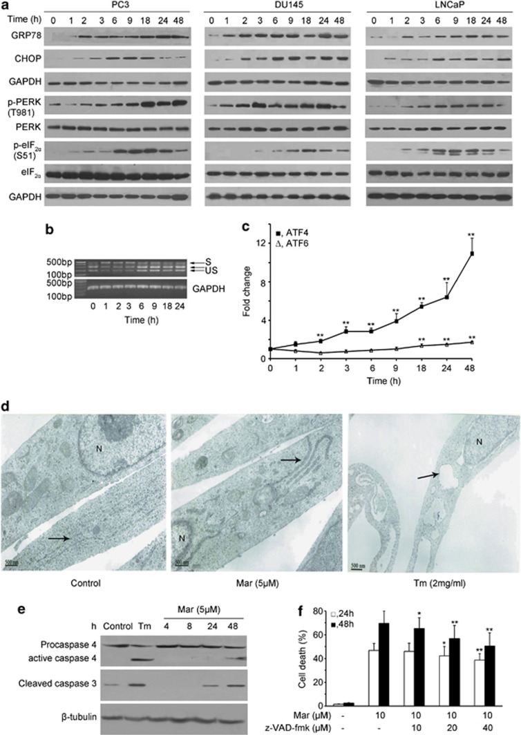
Figure 3.
Mar triggers ER stress in PCa cells. (a) Analysis of the effect of Mar on proteins associated with ER stress by western blotting. (b) Analysis of XBP1 splicing after Mar (10 μM) treatment in PC3 cells by RT-PCR. S, spliced XBP1; US, unspliced XBP1. (c) Measurement of ATF4 and ATF6 mRNA expressions in PC3 cells exposed to Mar by real-time RT-PCR. **P<0.01, compared with the control (0 h treatment). Data shown are means±S.D., n=3. (d) Changes of ER in cells treated with Mar (5 μM) using a transmission electron microscope. Tm (2 mg/ml) was taken as a positive control. The ER is indicated by black arrows. N, nucleus. Bar, 500 nm. (e) Analysis of caspase-4 and caspase-3 activation in Mar-treated cells by western blotting. (f) Effects of the apoptosis inhibitor z-VAD-fmk on Mar-induced cell death in PC3 cells. PC3 cells were incubated with z-VAD-fmk for 2 h prior to Mar treatment. Cell death was assayed by PI exclusion; *P<0.05, **P<0.01. Comparison between groups treated with Mar plus z-VAD-fmk or Mar alone. Data shown are means±S.D., n=3

