Figure 5.
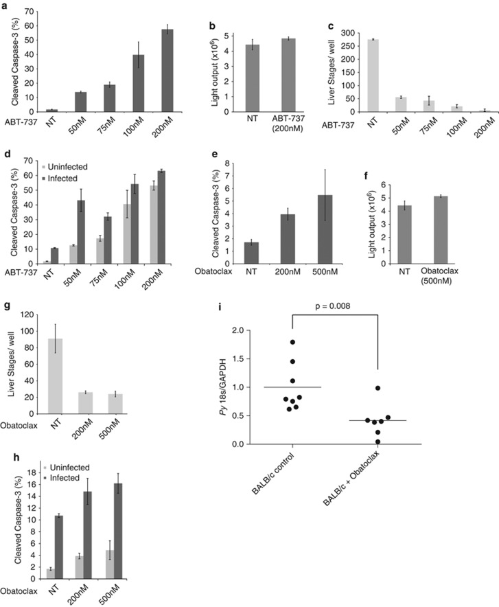
Bcl-2 family-targeted drugs eliminate LS-infected hepatocytes in vitro and in vivo via apoptosis. (a) ABT-737 induces apoptosis in Hepa 1-6 cells in a dose–response manner. Cells were treated with the indicated concentration of ABT-737 for 24 h. (b) Transgenic P. falciparum parasites that express a GFP-luciferase fusion were used to monitor growth in asexual blood stage culture. There is no difference between the signal obtained from drug-treated and untreated parasites. Parasites were synchronized as ring stages and allowed to develop for 48 h. Drug was replaced daily. (c) ABT-737 robustly eliminates P. yoelii parasite-infected host cells in vitro. LSs were visualized by microscopy and quantified by manual counting. Cells were treated with the indicated concentration of ABT-737 for 24 h, beginning 90 min after infection. (d) LS-infected host cells are more susceptible to ABT-737-mediated apoptosis than uninfected cells. Cells were treated with indicated concentrations of ABT-737 for 24 h, beginning 90 min after infection. Populations of caspase-3-positive cells were examined in infected and uninfected populations by flow cytometry. (e) Obatoclax induces apoptosis in Hepa 1-6 cells. Cells were treated with indicated concentrations of obatoclax for 24 h. (f) Transgenic P. falciparum parasites that express GFP-luciferase fusion were used to monitor growth in asexual blood stage culture. There is no significant difference between the signal obtained from drug-treated and untreated parasites. (g) Obatoclax eliminates P. yoelii LS-infected host cells in vitro. LSs were visualized by microscopy and quantified by manual counting. (h) Infected cells are more susceptible to obatoclax-mediated apoptosis than uninfected cells. Cells were treated with indicated concentrations of obatoclax for 24 h, beginning 90 min after infection. Populations of caspase-3-positive cells were examined in infected and uninfected populations by flow cytometry. (i) Obatoclax effectively eliminates LS-infected hepatocytes in vivo. BALB/cJ mice were treated with 5 mg/kg obatoclax once daily and infected with 100 000 P. yoelii sporozoites. LS burden was monitored by quantitative PCR 42–44 h after infection

