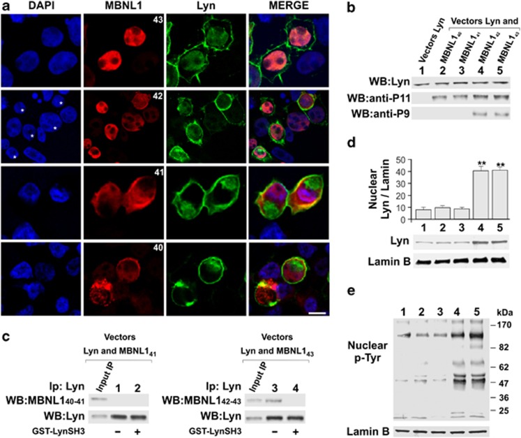
Figure 4.
Translocation of Lyn to the nuclei of 293T cells occurs only upon co-expression with MBNL142–43. (a) Representative confocal images of 293T cells co-transfected with plasmids carrying different MBNL1 isoforms (MBNL140–41–42–43 in pEF1-Myc/HisA vector) and Lyn (pCMV6-XL4/Lyn). Nuclei were labelled with DAPI (blue), MBNL1 with anti-P9 (MBNL142–43) or anti-P11 (MBNL140–41) antibodies (red) coupled to immunodetection of Lyn (green). Scale bar 10 μm. (b) Representative WB analysis of whole-cell lysates from 293T cells transfected with expression vectors for Lyn, MBNL140, MBNL141, MBNL142 and MBNL143 probed with anti-Lyn antibody (top strip), anti-P11 antibody (middle strip) and anti-P9 antibody (bottom strip). (c) Representative co-immunoprecipitation of four-fifths of whole-cell lysates from 293T cells, overexpressing MBNL141 (pEF1/MBNL141 (left panel), lanes 1 and 2), or MBNL143 (pEF1/MBNL143 (right panel), lanes 3 and 4) and Lyn (pCMV6-XL4/Lyn, lanes 1–4). Thirty-six hours after transfection, whole-cell lysates were immunoprecipitated with anti-Lyn in the absence (lanes 1 and 3) or presence (lanes 2 and 4) of the GST-Lyn SH3 domain. The immunocomplexes and one-fifth of the whole-cell lysates underwent WB analysis with anti-P11 or anti-P9 antibodies, respectively, and subsequently with anti-Lyn antibodies. The figure represents one of three independent experiments. The same set of experiments conducted with MBNL140 and MBNL142 isoforms demonstrated that these two proteins behave similarly to MBNL141 and MBNL143, respectively, again supporting the hypothesis that it is exon 5 that enables MBNL1s to interact with SFKs (data not shown). (d) Representative WB analysis of nuclear lysates from 293T cells transfected with expression vectors for Lyn (lanes 1–5), MBNL140 (lane 2), MBNL141 (lane 3), MBNL142 (lane 4) and MBNL143 (lane 5). The membranes were probed with anti-Lyn and with anti-Lamin B antibody as loading control. The resulting bands underwent densitometric analysis. The values (arbitrary units) represent the mean±S.D. of at least three experiments. Reference unit: lane 1 sample (only transfected with Lyn). **Significant difference for P<0.01 by Kruskal–Wallis analysis. (e) Representative WB analysis of nuclear lysates from 293T cells, transfected with expression vectors for Lyn (lanes 1–5), MBNL140 (lane 2), MBNL141 (lane 3), MBNL142 (lane 4) and MBNL143 (lane 5) and probed with anti-p-Tyr antibody. The membranes were reprobed with anti-Lamin B antibody as loading control

