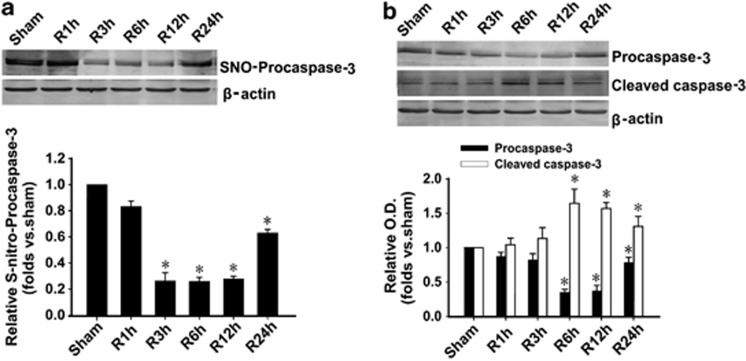
Figure 1.
Procaspase-3 is denitrosylated and activated during I/R in hippocampus. (a) Time course analysis of denitrosylation of procaspase-3 levels in hippocampal CA1 derived from sham-treated rats or rats with 15 min ischemia at various time points (1, 3, 6, 12, and 24 h) after reperfusion (R). n=4; *P<0.05 compare with sham group. (b) Time course analysis of procaspase-3 activation (cleaved caspase-3) levels in hippocampal CA1 derived from sham-treated rats or rats with 15 min ischemia at various time points (1, 3, 6, 12, and 24 h) after reperfusion. The samples were processed using the biotin switch method followed by western blotting. Antibodies aginst procaspase-3 and cleaved caspase-3 were used to measure the expression of procaspase-3 and its cleaved large part (17/19 kDa; n=4; *P<0.05 compare with corresponding sham group). Data are represented as means±S.D.

