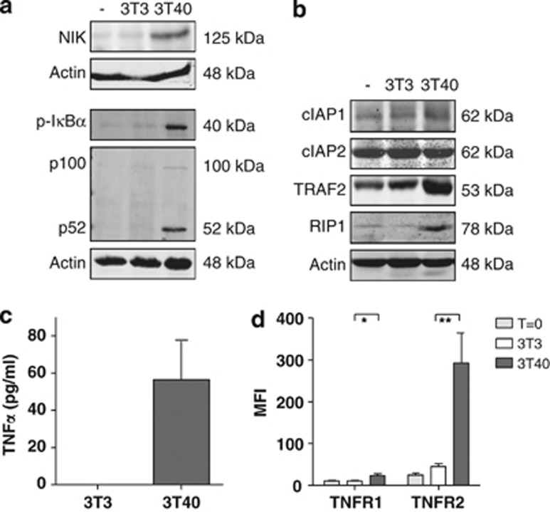
Figure 1.
Patient CLL cells stimulated with CD40L activate the non-canonical NF-κB pathway and the production of TNF, TNFR1 and TNFR2. (a) CLL cells were not cultured (−) or co-cultured for 48 h with 3T3 control feeder layer cells (3T3) or on CD40L-expressing 3T3 feeder layer cells (3T40) for 48 h. Lysates separated on SDS-PAGE gels and western blotted with the indicated antibodies, where actin was served as a loading control. Results of one representative CLL patient, of a total of nine analyzed, are shown. (b) CLL cells were treated as in a. Protein levels of cIAP1, cIAP2, TRAF2, TRAF6 and RIPK1 were analyzed by immunoblotting on total lysates. Blots from one representative CLL patient, of a total of three analyzed, are shown. (c) CLL cells were co-cultured with 3T3 or 3T40 cells for 72 h, after which supernatants were collected and TNFα levels were measured using ELISA. The means and S.E.M. of n=16 independent CLL samples are represented. (d) Expression levels of TNFR1 and TNFR2 were measured by flow cytometry of freshly isolated, 3T3 and 3T40 co-cultured CLL cells (n=8, independent CLL samples). Error bars represent S.E.M. Asterisks indicate statistical significance: *0.01<P<0.05 and **0.001<P<0.01

