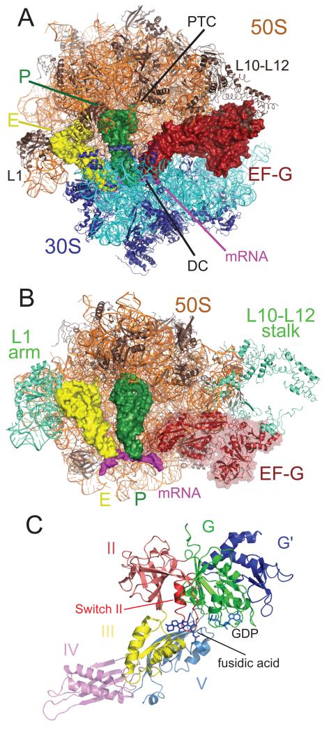
Figure 2.
EF-G in the post-translocational state of the ribosome. A. Overall view of EF-G in the ribosome. EF-G is shown in reddish-brown, the 50S subunit on top is shown in orange, the 30S subunit below is shown in cyan, P-site tRNA in green, and E-site tRNA in yellow and the mRNA in purple. The decoding center (DC) in the 30S subunit and the peptidyl transferase center (PTC), and the L1 and L10-L12 stalks are indicated as shown.B. Global changes in the 50S subunit as a result of EF-G binding. The mobile regions of the 50S subunit are indicated in teal, and include the L1 stalk (both RNA and protein), the L11 region, and the the L10-L12 stalk. C.The structure of EF-G bound to fusidic acid and GDP in the ribosome. The various domains of EF-G are colored as shown.

