Figure 1.
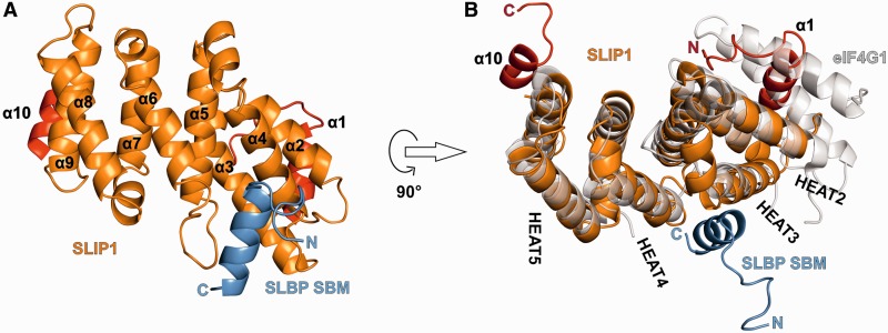
Structure of SLIP1 bound to the SBM of SLBP. (A) Structure of the D.r. SLIP1–SLBP complex. SLBP is in blue. SLIP1 is shown with the 4 HEAT repeats in orange and the N- and C-terminal helices in red. The N- and C-termini of SLBP are labeled and the helices of SLIP1 are numbered. (B) The structure of D.r. SLIP1–SLBP is shown in an orientation related by a 90° rotation around a horizontal axis with respect to panel A. It is superposed on the MIF4G domain of eIF4G (in gray, PDB code 2VSO). The HEAT repeats of SLIP1 are numbered.

