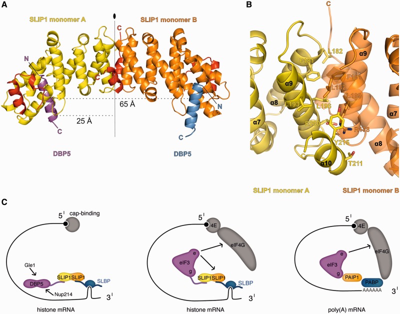
Figure 6.
Concurrent binding of two SBMs to a SLIP1 homodimer. (A) Structure of the dimeric SLIP1–SLBP complex observed in the crystal lattice. The two monomers (A and B) are labeled. The vertical line indicates the 2-fold symmetry of the dimer. (B) Close-up view of the dimerization interface, shown in the same orientation as in panel A. Selected conserved residues are shown in stick representation and labeled. For clarity, helix 8 is shown with 50% transparency. (C) Left panel: model for the involvement of SLBP in histone mRNA export. The SLIP1 heterodimer is shown in yellow and orange, as in panel A. The model does not include additional levels of regulation, such as phosphorylation (39). Central panel: model for the mechanisms of translation stimulation of histone mRNAs as compared with that of poly(A)-containing mRNAs (right panel). Arrows indicate additional interactions reported in the literature.

